The procedure has taken years to develop, is difficult for surgeons to master and yet could not be simpler.
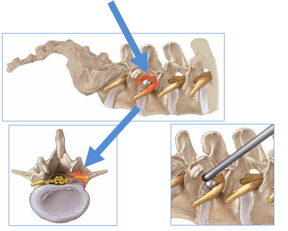
Using X-ray guidance, a probe, the size of a pencil, is passed through the natural opening in the side of the spine through which the nerve root at the affected level exits - the nerve root foramen.
Then, aided by the huge magnification of the endoscope, its bright illumination and high resolution screens, the disc prolapse or narrowed canal is gradually and gently cleared until the nerve can be seen free in the canal.
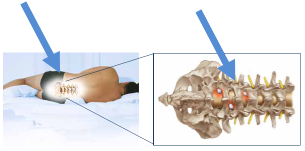
The probe is well tolerated with local anaesthetic and some sedation so general anaesthesia is not required. Indeed, it is preferable to be able to observe the nerve’s function throughout the procedure. Typically the patient lies on their side slumbering to be woken occasionally by us, asking them to move their foot or wiggle their toes.
It is often over in about an hour though there is never a hurry.
For a more detailed explanation of the techniques and benefits of endoscopic spinal surgery, please click here.
How is endoscopic disc surgery done?
The procedure is carried out with the patient on their side under heavy sedation but without a full general anaesthetic. The procedure is very well tolerated and the fact it does not require a general anaesthetic makes it safer for both the nerve root and the patient.
The endoscopic tube is guided into the spinal canal through a natural opening in the side of the spinal canal, the foramen, below the exiting nerve root. This gives direct access to the prolapsed disc and the nerves it is compressing.
You are lying on your side during the operation with the bad leg uppermost. You have the anaesthetist with you all the time. Their job is to keep enough pain relief and sedation going to ensure you are comfortable and with today’s drugs they are very good at it.
A small incision is made for the endoscope and this is guided by X-rays screening into the opening. Once in position the disc fragments are removed. Your nerves are monitored all the time to ensure they are not injured.
After that the job is done. A single stitch is put in the skin and you return to your room. You may get up that day. Home time is usually the next day.
THE EQUIPMENT
Like most endoscopic equipment, it looks more complicated than it is. A fibre optic cable takes light to the probe and returns images to the camera. The surgeon operates looking between the screens on the camera stack and the X-ray machine.
The system employed at The Spine Surgery London is the TESSYS system made by Joimax. We believe this to be by far the most advanced spinal endoscopic system available. The equipment is of course still hugely expensive though we believe the investment made by our supporting hospital, The Princess Grace Hospital, will see our patients benefit immensely. Less operating time, tissue damage, recovery time and less time in hospital, off work, in physiotherapy and in pain.